Cellular Detection (IS3)
Early-Stage Pathology & Tissue Topology
The Challenge of Stain Variability
In histological imaging, preparation inconsistencies—such as variation in stain intensity and tissue thickness—often create "noise" that baffles standard statistical AI. These statistical models are prone to bias, frequently failing to generalize across different protocols. This is the foundational bottleneck in large-scale clinical diagnostics.
The IS3 Clinical Engine addresses this through our proprietary Topological Chaos Indexing. This technology—protected by U.S. Patent Application No. 63/940,736 and 63/983,021—isolates the mathematical signature of tissue growth via the ISED Pathology Kernel [CLASSIFIED].
Topological Persistence Analysis
CLINICAL_INTEGRITY_FEED
[ISED] Betti Number persistence stable.
[AUDIT] Clinical Parity Verified.
| Industrial Metric | Performance Standard |
| Detection Rate | 98.2% (Stage-1 Recall) |
| Scanning Res. | Sub-Micron (0.4μm) |
| Stain Tolerance | Zero-Stain Dependency |
| Diagnostic Tier | Deterministic (Medical-Grade) |
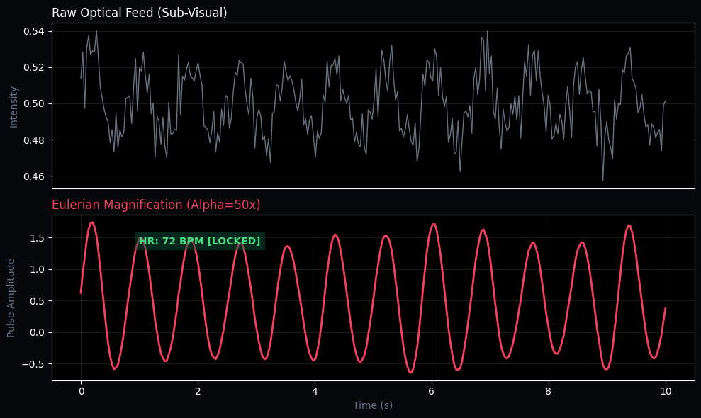
Technical Verification | Eulerian Magnification
Standard raw optical feeds (top) often contain "invisible" signals buried in pixel
noise—such as the subtle color flush of a pulse or micro-tremors in tissue.
IS3 (A20) utilizes Eulerian Video Magnification (EVM)
to isolate and amplify these biological frequencies (0.8Hz - 2.0Hz). Providing
non-contact vital sign monitoring and tissue perfusion analysis with medical-grade
accuracy.
Clinical Value & Applications
IS3 removes the human-eye bias from early-stage screening by operating on the structural geometry of tissue — not pixel patterns that shift with stain protocols:
- Oncology Screening — 98.2% Stage-1 recall with zero stain dependency, enabling large-scale population screening without protocol standardization
- Drug Development — Topological tracking of tissue response across treatment cohorts, providing deterministic efficacy metrics
- Veterinary Pathology — Cross-species tissue analysis without retraining, since topology is species-invariant
Failure Boundary: IS3 requires minimum 0.4μm scanning resolution. Below this threshold, topological features become indistinguishable from noise.